Article
주걱댕강나무(Diabelia spathulata)의 체세포배발생 및 식물체 재분화 특성
김도연1,2,
박지은3,
변하연3,
강준원4,*
Somatic Embryogenesis and Plantlet Regeneration in Diabelia spathulata
Do Yeon Kim1,2,
Ji Eun Park3,
Ha Yeon Byun3,
Jun-Won Kang4,*
Author Information & Copyright ▼
1Department of Forestry, Kyungpook National University, Daegu 41566, Korea
2Department of Agriculture, Forestry and Bioresources, Seoul National University, Seoul 08826, Korea
3National Institute of Biological Resources, Miryang 50452, Korea
4School of Forest Sciences and Landscape Architecture, Kyungpook National University, Daegu 41566, Korea
© Copyright 2026 Korean Society of Forest Science. This is an Open-Access article distributed under the terms of the
Creative Commons Attribution Non-Commercial License (http://creativecommons.org/licenses/by-nc/4.0/) which permits
unrestricted non-commercial use, distribution, and reproduction in any
medium, provided the original work is properly cited.
Received: Jun 16, 2025; Revised: Dec 22, 2025; Accepted: Mar 10, 2026
Published Online: Mar 31, 2026
요 약
주걱댕강나무(Diabelia spathulata)는 경상남도 양산시 천성산에서 제한적으로 분포하는 희귀수종으로 보존 연구를 위해 효율적인 증식 기술 확립이 필요하다. 본 연구는 주걱댕강나무 엽절편을 이용하여 체세포배 발생을 유도하고, 배양 조건이 체세포배 형성에 미치는 영향을 평가하고자 수행하였다.
엽절편을 다양한 농도의 BAP와 NAA가 첨가된 MS 기본배지에서 배양한 결과, 모든 처리에서 캘러스가 유도되었고, 일부 처리구에서는 기관 형성이 관찰되었다. 최적 조건으로는 BAP 1.0 mg/L + NAA 2.0 mg/L 처리구였으며 직경 16.67 mm, 생중량 295.8 mg의 캘러스가 형성되었다. 배발생 캘러스 및 체세포배 형성은 낮은 무기염 농도(1/4~1/2 MS)와 3% sucrose 첨가 조건에서 가장 활발하게 발생되었으며, 배양 4~8주 후 구형, 심장형, 어뢰형 단계를 거쳐 자엽기 단계로 발달하였다. 활성탄 첨가는 0.01% 농도에서 신초 출현율을 증가시켰으나, 0.2% 농도에서는 발달을 억제하였다.
이러한 결과는 주걱댕강나무에서 체세포배를 통한 식물체 재분화 체계를 확립할 수 있음을 보여주며, 향후 희귀 수종의 대량증식 및 복원 연구에 활용될 수 있을 것으로 판단된다.
Abstract
Diabelia spathulata is a rare plant species endemic to Cheonseongsan Mountain, Yangsan-si, Gyeongsangnam-do, Korea; thus, establishing efficient propagation techniques is critical for its conservation. This study aimed to induce somatic embryogenesis from leaf explants of D. spathulata and evaluate the effects of culture conditions on somatic embryo formation.
Leaf explants cultured on Murashige and Skoog (MS) medium supplemented with various concentrations of 6-benzylaminopurine (BAP) and naphthaleneacetic acid (NAA) produced callus under all treatment conditions, with occasional organogenesis observed under low growth regulator concentrations. Optimal callus formation was achieved under treatment conditions of 1.0 mg/L BAP and 2.0 mg/L NAA, which yielded callus with a diameter of 16.67 mm and a fresh weight of 295.8 mg. Embryogenic callus and somatic embryo formation were most active on a low-salt medium (1/4-1/2 MS) with 3% sucrose, developing through the globular, heart, and torpedo stages to the cotyledonary stage within 4-8 weeks. Activated charcoal at 0.01% increased shoot emergence, whereas 0.2% inhibited further development.
These findings provide fundamental information on the somatic embryogenesis of D. spathulata and establish a reliable basis for the mass propagation and conservation of this rare species.
Keywords: Diabelia spathulata; leaf culture; somatic embryogenesis; callus induction; embryogenic callus; plant regeneration
서 론
주걱댕강나무(Diabelia spathulata)는 인동과(Caprifoliaceae)에 속하는 낙엽 활엽 관목으로, 5월경 주걱 모양의 황백색 꽃이 피어 관상적 가치가 높은 식물이다. 국내에서는 경상남도 양산시 천성산 일대에서만 자생지가 확인되었으며, 전체 점유 면적은 4 km2 미만으로, 국제자연보전연맹(IUCN) 적색목록에서 취약(Vulnerable) 등급으로 분류된 희귀식물이다(KNA, 2021).
기존에는 일본 특산식물로 알려졌으나, 2003년 천성산에서 자생지가 발견되면서 한국 고유 식물로서의 학술적⋅보전적 가치가 부각되었다(Yi et al., 2021). 주걱댕강나무는 뿌리줄기를 통한 영양번식으로 군락을 유지하며(Jang, 2021), 결실률 또한 매우 낮아 유전적 다양성이 제한될 가능성이 제기되고 있다(Kim and Kim, 2018). 자생지는 등산, 채집 등 인위적 간섭에 노출되어 있으며, 병해충과 환경변화에도 취약한 상태이다. 기후변화 또한 생물다양성 손실과 희귀식물의 생존 가능성을 저하시킬 수 있는 주요 요인으로 지적된다(Jeong et al., 2010).
이러한 배경에서 현지 외(ex situ) 보전을 수행할 수 있는 기내 대량증식 조건 확립은 매우 중요하다. 식물 조직배양 기술은 자생지를 교란하지 않고 대량증식이 가능하다는 점에서 희귀식물 보전 수단으로 주목받고 있다. 국내에서도 문관과(Ahn and Lee, 2023), 회화나무(Im et al., 2024) 등 자원식물뿐 아니라, 개느삼(Moon et al., 2008), 두메닥나무(Chu et al., 2020), 미선나무(Moon et al., 1999), 좀개미취(Park et al., 2024) 등 희귀 자생 식물을 대상으로 조직배양 기반 증식 기술이 활발히 개발되고 있다.
한편, 댕강나무속(Diabelia) 식물에 대해서는 댕강나무와 꽃댕강나무의 삽목 증식 연구(Kim, 1996; Dirr, 1977; Ahn, 2003)가 보고되었으나, 조직배양을 활용한 대량증식 기술 개발은 아직 보고된 바 없다. 주걱댕강나무 역시 종자 및 삽목 증식 연구(Jang, 2021)는 수행되었으나, 조직배양 기반 체세포배발생 연구는 미흡한 실정이다.
따라서 본 연구에서는 주걱댕강나무의 엽절편을 이용한 엽배양 기술을 바탕으로, 생장조절제 조합, 광조건, 무기염 및 탄소원 농도 등 배양 조건이 캘러스 유도 및 체세포배 형성에 미치는 영향을 평가하고, 기내 증식 가능성을 체계적으로 분석하고자 하였다.
재료 및 방법
1. 식물재료 및 배양 조건
본 연구에 사용된 주걱댕강나무[Diabelia spathulata (Siebold & Zucc.) Landrein]는 경상남도 양산시 천성산 일대에서 채집하였으며, 기내에서 삽목 후 2개월 동안 생육하였다. 이후 삽목한 식물체에서 잎을 채취하여 75% 에탄올에 2분 처리 후, 멸균수로 2회 수세하였다. 이어 0.5% 차아염소산나트륨(sodium hypochlorite, NaOCl)에서 10분 침지 후 멸균수로 3회 수세하여 표면소독하였다. 이후 0.5 cm × 0.5 cm의 크기로 절편체를 제작하였으며, 각 실험구에 5개의 절편을 배치하고 3회 반복 수행하여 배지에 치상하였다.
실험에 사용한 모든 배지는 MS 배지(Murashige and Skoog, 1962)에, 30 g/L sucrose와 8 g/L plant agar를 첨가하고, pH를 5.8로 조정하였다. 이후 1.5 기압, 121 °C 조건에서 15분간 멸균 후 Petri-dish에 40 mL씩 분주하였다. 배양조건은 25±2 °C에서 유지하였고, 광조건은 2,000 lux의 광에서 16시간 노출, 암조건은 24시간 동안 빛을 차단한 조건에서 배양하였다. 4주마다 동일 배지로 계대 배양하였으며, 12주 후 각 실험구의 캘러스 유도율, 배발생 캘러스 유도율, 기관 분화 유도율을 조사하였다.
통계 분석은 SPSS(version 26.0)를 이용하여 일원 배치 분산분석(one-way ANOVA)과 Duncan's Multiple Range Test (DMRT; Duncan, 1955)로 유의수준 5%(p≤0.05)에서 검증하였다. 모든 결과는 평균±표준오차(means±SE)로 나타냈다.
2. BAP와 NAA의 농도 조합에 따른 캘러스 유도 및 생장 조건 최적화
캘러스 유도에 가장 적합한 식물생장조절제의 종류와 농도를 평가하기 위해, 사이토키닌 계열 식물생장조절제(BAP, Kinetin, Zeatin)와 옥신계열 식물생장조절제(2,4-D, NAA)를 혼용 처리한 배지에 엽절편을 치상하였다.
사이토키닌 계열 식물생장조절제 종류에 따른 캘러스 유도를 비교하기 위해 BAP, Kinetin, Zeatin을 1.0 mg/L로 고정하고, NAA를 농도별(1.0, 2.0, 3.0 mg/L)로 혼용 처리하였다. 또한, 옥신 계열 식물생장조절제 종류에 따른 캘러스 유도를 비교하고자 BAP 0.5 mg/L에 NAA 또는 2,4-D를 각각 농도별(1.0, 2.0, 4.0 mg/L)로 혼용 처리하여 실험을 진행하였다. 한편, 식물생장조절제의 고농도 처리에 따른 반응을 확인하기 위해 BAP와 NAA를 5.0 mg/L 이상의 농도로 처리하는 실험을 병행하였다.
3. 배지 농도에 따른 배발생캘러스의 유도
배발생 캘러스(Embryogenic callus, EC)의 유도를 위해, 잎에서 유도한 캘러스를 직경 2 mm로 절단하여 식물생장조절제를 처리하지 않은 배지(1/4 MS, 1/2 MS, MS)와 BAP 1.0 mg/L를 단용 처리한 1/2 MS, MS 배지에 각 5개씩 3회 반복으로 치상한 후 광조건에서 배양하였다. 이후 배발생 캘러스 유도율과 증식 과정을 관찰하였다.
4. 배지 농도 및 Sucrose 농도에 따른 체세포배 형성 비교
배발생 캘러스로부터 체세포배 형성에 적합한 배지 농도와 sucrose 농도를 평가하기 위해, 무기염 농도를 1/4배, 1/2배, 1배, 2배로 조정한 MS 배지와 sucrose 농도를 0, 1, 3, 5, 7%로 조절한 1/2 MS 배지를 제작하였다. 이후 각 실험구에 직경 2 mm의 배발생 캘러스를 5개씩 3회 반복 치상하였으며, 광조건에서 5주간 배양한 후, 발생한 정상적인 체세포배 개수를 조사하였다.
정상적인 체세포배는 구형으로 형성되고, 세포벽이 균일하며 본엽이 발달할 수 있는 상태로 정의하였다(Ammirato, 1983; Williams and Maheswaran, 1986).
5. 활성탄 처리에 따른 식물체 재분화 비교
활성탄 처리에 따른 체세포배의 재분화율을 평가하기 위해, MS 기본배지에 활성탄을 0, 0.01, 0.02, 0.1, 0.2%로 첨가하였다. 각 실험구에는 자엽 2개를 가진 체세포배를 5개씩 3회 반복 치상하였다. 이후 광 조건에서 배양하였으며, 3주마다 동일 배지로 계대 배양하였다. 6주 후 본엽이 나온 개체를 대상으로, 체세포배당 출현한 신초의 수를 조사하였다.
결과 및 고찰
1. 캘러스 유도 및 증식
1) 식물생장조절제 혼용 처리에 따른 캘러스 발생
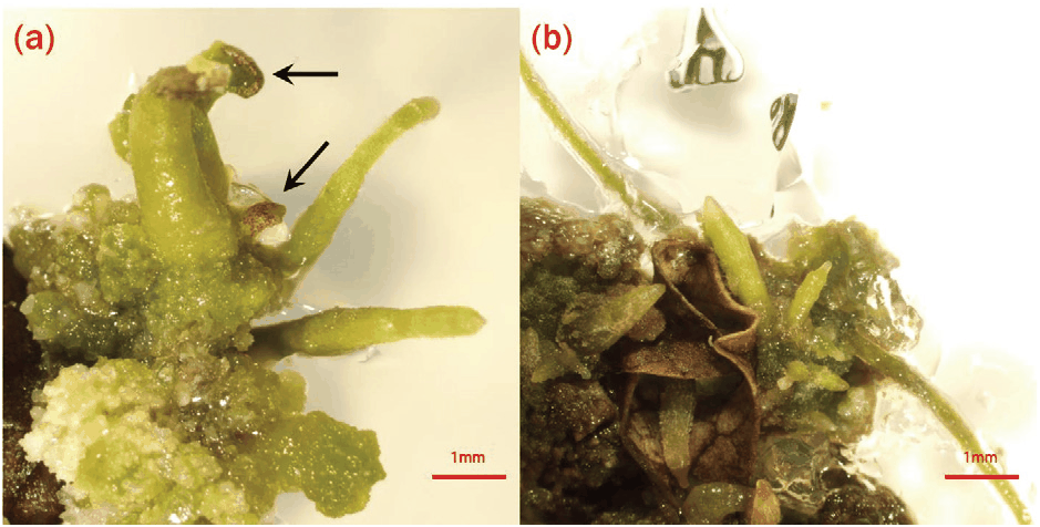
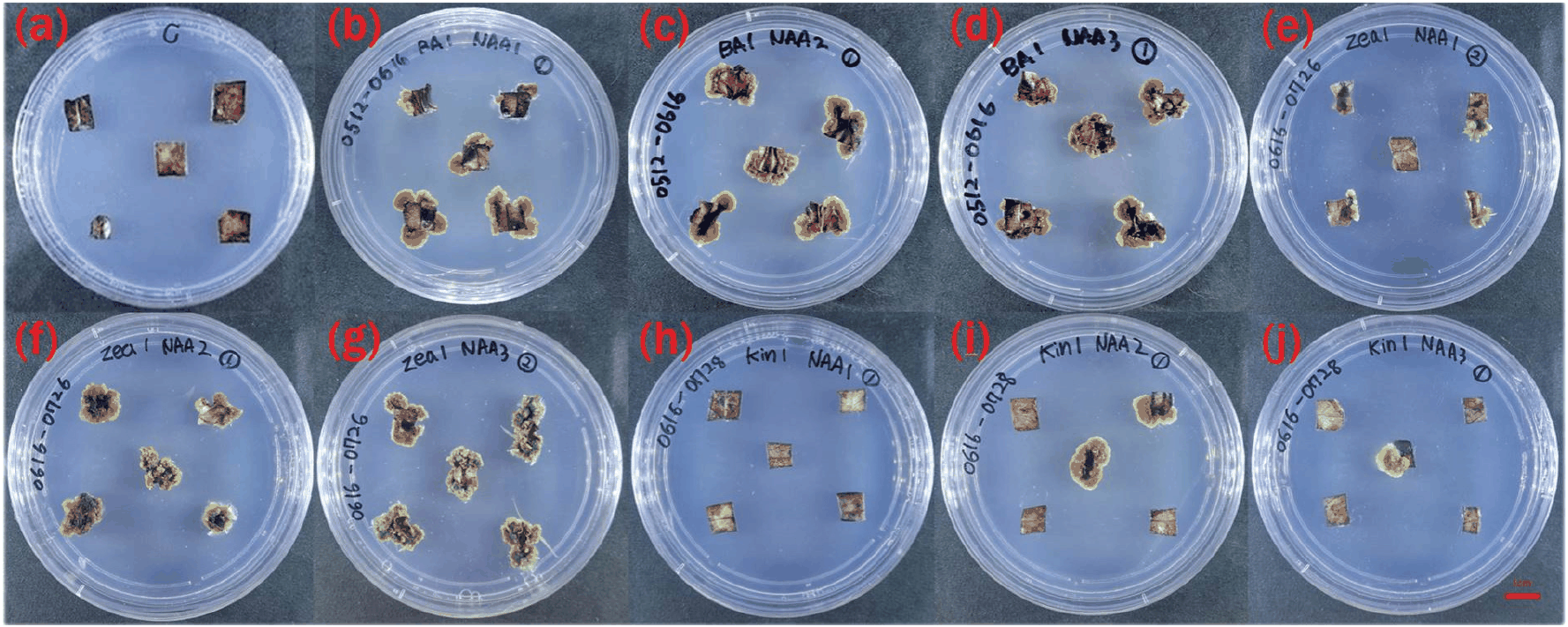
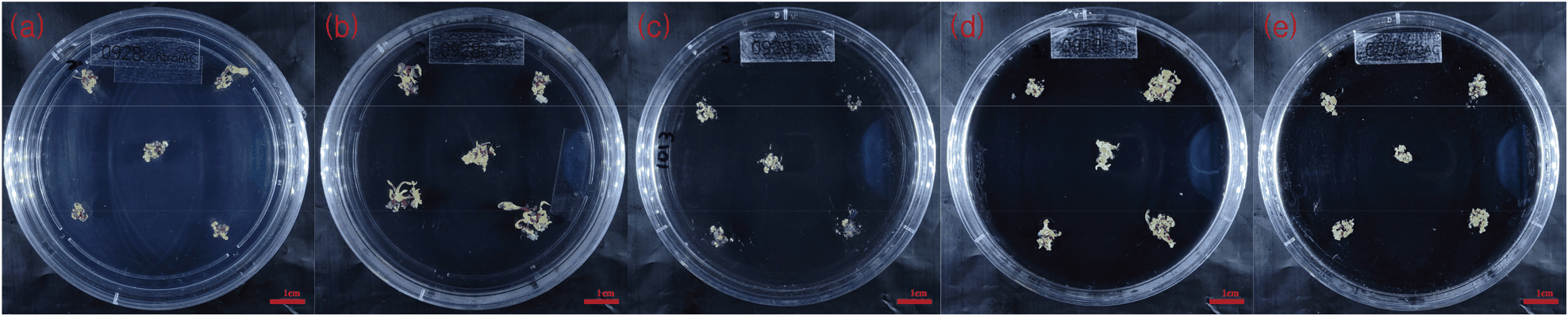
본 연구에서는 주걱댕강나무 캘러스 유도에 대한 식물생장조절제의 영향을 평가하였다. 치상 8주 후, 모든 처리구에서 비배발생성(non-embryogenic) 캘러스가 형성되었으며, 2,4-D 단용 처리구에서는 캘러스 유도 및 증식 속도가 현저히 낮았다(데이터 미제시). 일반적으로 식물 조직배양에서 auxin과 cytokinin의 혼용 처리는 세포 분열을 촉진하여 캘러스 형성을 증가시키는 경향이 있다. 특히 BAP 처리구에서 캘러스 유도율은 73.3~100%로 나타났으며(Table 1), 이에 따라 BAP와 NAA 농도별 혼용 처리 실험을 추가 수행하였다. 12주 계대배양 후, 일부 처리구에서 일시적인 기관 분화가 관찰되었다(Figure 1). 이는 체세포 유도 과정 중 발생한 비특이적 반응으로 판단된다. 일원 배치 분산분석(one-way ANOVA) 결과, 캘러스의 직경과 생중량에서 유의미한 차이가 관찰되었으며, 특히 BAP 1.0 mg/L + NAA 2.0 mg/L 조합에서 가장 뚜렷한 증가가 확인되었다(Table 2). Zeatin과 NAA 혼용 처리구에서 캘러스 생중량이 가장 크게 나타났으며, zeatin 처리는 비특이적 부정근 형성이 뚜렷하였다(Figure 2). 이러한 반응은 체세포배 유도와 직접적인 관련이 없는 부수적 생리 반응으로 해석된다. 그러나 모든 처리에서 배발생 캘러스는 유도되지 않았다.
Table 1.
Effect of the concentration of cytokinins (BAP, zeatin, kinetin) and auxin (NAA) added to MS on callus induction from leaf segments after 8 weeks of culturez.
| Growth regulators (mg/L) |
No. of leaf segments cultured |
No. of leaf segments produced callus |
% of callus formation |
| BAP |
zeatin |
kinetin |
NAA |
| 0.0 |
0.0 |
0.0 |
0.0 |
15 |
0 |
0 |
| 1.0 |
|
|
1.0 |
15 |
11 |
73.3 |
| 1.0 |
|
|
2.0 |
15 |
15 |
100.0 |
| 1.0 |
|
|
3.0 |
15 |
13 |
86.6 |
|
|
1.0 |
|
1.0 |
15 |
7 |
46.6 |
|
|
1.0 |
|
2.0 |
15 |
9 |
56.6 |
|
|
1.0 |
|
3.0 |
15 |
10 |
66.6 |
|
|
|
1.0 |
1.0 |
15 |
1 |
3.3 |
|
|
|
1.0 |
2.0 |
15 |
2 |
6.6 |
|
|
|
1.0 |
3.0 |
15 |
1 |
3.3 |
Download Excel Table
Figure 1.
Organogenesis in cultured leaf explants of D. spathulata : (a) Indirect shoot organogenesis from callus of Diabelia leaf segments on the combination BAP and NAA after 4 weeks in culture. (b) Indirect root organogenesis from callus of Diabelia leaf segments on the combination zeatin and NAA after 4 weeks in culture. Scale bars represent 1 mm.
Download Original Figure
Table 2.
Effect of the concentration of cytokinins (BAP, zeatin, kinetin) and auxin (NAA) added to MS on callus induction from leaf segments after 12 weeks of culturez.
| Growth regulators (mg/L) |
No. of leaf segments cultured |
No. of leaf segments produced callus |
% callus induction |
% organogenesis induction |
Length size of callus(mm)y |
Fresh weight (mg) of callus/explantx |
| BAP |
zeatin |
kinetin |
NAA |
| 0.0 |
0.0 |
0.0 |
0.0 |
15 |
0 |
- |
- |
0.0±0.0 |
0.0±0.0 |
| 1.0 |
|
|
1.0 |
15 |
12 |
80 |
- |
14.67±0.95 |
180.4±40.83 |
| 1.0 |
|
|
2.0 |
15 |
15 |
100 |
6.7 |
16.67±0.94* |
295.8±45.53* |
| 1.0 |
|
|
3.0 |
15 |
13 |
86.7 |
- |
14.36±0.77 |
149.8±38.08 |
|
|
1.0 |
|
1.0 |
15 |
10 |
66.7 |
26.7 |
11.32±0.18 |
182.4±66.14 |
|
|
1.0 |
|
2.0 |
15 |
15 |
100 |
33.3 |
13.27±0.74 |
183.73±33.45 |
|
|
1.0 |
|
3.0 |
15 |
12 |
80 |
53.3 |
14.15±0.30 |
293.73±60.8 |
|
|
|
1.0 |
1.0 |
15 |
1 |
6.7 |
- |
9.08±0.94 |
18.06±1.2 |
|
|
|
1.0 |
2.0 |
15 |
2 |
13.3 |
- |
9.84±0.94 |
64.93±29.72 |
|
|
|
1.0 |
3.0 |
15 |
1 |
6.7 |
- |
9.57±0.98 |
33.2±14.26 |
Download Excel Table
Figure 2.
Callus induction from leaf segments of D. spathulata on MS containing cytokinins (BAP, zeatin, kinetin) and auxin (NAA) after 12 weeks of culture. Scale bars represent 1 cm.
(a) 0.0 mg/L (control) (b) BAP 1.0 mg/L + NAA 1.0 mg/L (c) BAP 1.0 mg/L + NAA 2.0 mg/L (d) BAP 1.0 mg/L + NAA 3.0 mg/L (e) zeatin 1.0 mg/L + NAA 1.0 mg/L (f) zeatin 1.0 mg/L + NAA 2.0 mg/L (g) zeatin 1.0 mg/L + NAA 3.0 mg/L (h) kinetin 1.0 mg/L + NAA 1.0 mg/L (i) kinetin 1.0 mg/L + NAA 2.0 mg/L (j) kinetin 1.0 mg/L + NAA 3.0 mg/L.
Download Original Figure
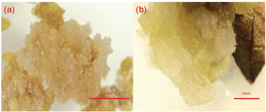
Auxin 종류별 비교 결과, BAP와 2,4-D 혼용 처리에서 캘러스 유도율은 86.7~100%로 상대적으로 높았으며, 캘러스 직경은 유의한 차이가 없었으나 생중량은 BAP 0.5 mg/L + 2,4-D 1.0 mg/L 처리구에서 가장 크게 증가하였다(데이터 미제시). 하지만 BAP와 2,4-D 조합으로 유도된 캘러스는 모두 비배발생성 조직으로 확인되었으며, 배발생 캘러스는 관찰되지 않았다[Figure 3(a)]. 이러한 결과는 다양한 식물 종에서 2,4-D가 배발생 캘러스 유도에 효과적이라는 기존 연구들과(Tahir et al., 2011; Malik et al., 2003; Phua et al., 2016; Dhir et al., 2014; Dalila et al., 2013; Pinto et al., 2002) 상반되는 결과이다[Figure 3(b)]. 또한, BAP + NAA 혼용 실험구의 캘러스는 연두색을 나타내며 생리적 안정성이 높았다. 이에 따라 본 연구에서는 캘러스 유도 및 계대배양에 적합한 Auxin으로 NAA를 선택하고, 최적 농도 조합 탐색을 위한 후속 실험을 설계하였다.
Figure 3.
Callus induced by BAP and 2,4-D. (a) Non-embryogenic callus (NEC) inoculated on MS medium supplemented with 0.5 mg/L BAP and 2.0 mg/L 2,4-D ; (b) Embryogenic callus (EC) inoculated on 1/2MS hormone free medium. Scale bars represent 1 mm.
Download Original Figure
2) BAP와 NAA 혼용 처리에 따른 캘러스 유도 및 증식 최적화
건강한 연두색 캘러스의 형성을 바탕으로, BAP와 NAA의 혼용 처리가 D. spathulata의 캘러스 유도에 가장 효과적인 조합으로 평가되었다. 이에 따라 두 생장조절제의 농도별 조합을 달리한 배지를 이용해 캘러스 유도율, 직경 및 생중량을 비교하였다.
BAP 및 NAA의 유효 농도 범위(0.5~2.0 mg/L) 내 혼용 처리구에서는 치상 4주 후부터 캘러스 유도가 시작되었으며, 8주차부터 직경과 생중량이 빠르게 증가하였다. 12주 차에는 BAP 0.5 mg/L + NAA 1.0 mg/L를 제외한 모든 처리구에서 66.7~100%의 높은 캘러스 유도율을 보였다. 직경 측정 결과, BAP 0.5 mg/L + NAA 2.0 mg/L 처리구에서 가장 넓은 직경(17.62 mm)이 기록되었고, 생중량은 BAP 1.0 mg/L + NAA 2.0 mg/L 처리구에서 가장 높은 수치(295.8 mg)를 나타냈다. 일원 배치 분산분석 결과, 직경과 생중량 모두에서 통계적으로 유의한 차이가 확인되었다(Table 3).
Table 3.
Effect of cytokinin (BAP) and auxin (NAA) added to MS on callus induction from leaf segments after 12 weeks of culturez.
| Growth regulators (mg/L) |
No. of leaf segments cultured |
No. of leaf segments produced callus |
% callus induction |
Length size of callus(mm)y |
Fresh weight (mg) of callus/explantx |
| BAP |
NAA |
| 0.0 |
0.0 |
15 |
0 |
- |
0.0±0.0 |
0.0±0.0 |
| 0.5 |
1.0 |
15 |
2 |
13.3 |
10.37±0.52 |
34±5.05 |
| 0.5 |
2.0 |
15 |
12 |
80 |
17.62±1.14* |
283.3±52.65 |
| 0.5 |
3.0 |
15 |
10 |
66.7 |
13.68±0.81 |
80±11.91 |
| 1.0 |
1.0 |
15 |
12 |
80 |
14.67±0.95 |
180.4±40.83 |
| 1.0 |
2.0 |
15 |
15 |
100 |
16.67±0.94 |
295.8±45.53* |
| 1.0 |
3.0 |
15 |
13 |
86.7 |
14.36±0.77 |
149.8±38.08 |
Download Excel Table
그러나 16주 이후 일부 캘러스에서 부정근 형성이 관찰되었으나, 이는 체세포배발생과 직접적인 관련이 없는 반응으로 판단되었다. 이러한 반응은 캘러스가 장기 배양되는 과정에서 생리적 불균형이나 세포 내 호르몬 농도 편차로 인해 나타나는 일시적 기관화 현상으로 해석된다. 비록 이러한 반응이 본 연구의 주 목적인 체세포배발생 과정과 직접적으로 연결되지는 않았으나, 주걱댕강나무 캘러스 조직이 적절한 자극 조건하에서 간접적 기관형성(indirect organogenesis)을 통해 재분화될 수 있는 전분화능(totipotency)을 갖추고 있음을 시사하는 지표로 판단된다. 그러나 이후 캘러스의 갈변 및 고사 현상이 점차 증가하면서 생장이 정체되었으며, 일부 조직은 생리적 노화에 따른 조직 퇴화도 함께 나타났다.
따라서 본 배지 조성은 캘러스의 초기 유도 및 증식에 최적화되어 있으나, 안정적인 체세포배 유도 및 발달 단계로의 전환을 도모하기 위해서는 배지 조성 및 생장조절제 비율의 정밀한 조정이 필요할 것으로 사료된다. 특히, 5.0 mg/L 이상의 고농도 처리구에서는 캘러스의 심한 갈변(Browning) 및 생장 억제가 관찰되어 분석 대상에서 제외하였으며(데이터 미제시), 본 연구에서는 재분화 효율이 높은 2.0 mg/L 이하의 적정 농도 처리 결과를 중심으로 체세포배 중심의 재분화 체계를 확립하고자 하였다. 또한, 배양 환경에 따른 캘러스 유도 효율을 확인하기 위해 광조건과 암조건을 비교한 결과, 모든 처리구에서 100%의 유도율을 보여 광조건에 따른 유의미한 차이는 없었다. 다만, 캘러스의 초기 증식 속도와 후속 배발생을 고려하여 이후 모든 실험은 광조건 하에서 수행하였다. 이에 따라, 확보된 캘러스로부터 효율적인 배발생 능력을 유도하고 체세포배로의 발달을 촉진하기 위하여 무기염 농도 및 탄소원 등을 조절한 배발생 캘러스 유도 배지에서의 배양 특성을 추가로 분석하였다.
2. 배발생 캘러스(Embryogenic callus; EC) 유도
1번 실험에서 최적 조건으로 유도된 녹색 캘러스에서 새로운 배지에 치상하여 관찰한 결과, 배양 8주 후 녹색 캘러스로부터 흰색 또는 노란색 배발생 캘러스가 혼재되거나, 노란색 배발생 캘러스가 단독으로 형성되는 현상이 관찰되었다. 형성된 노란색 배발생 캘러스를 동일한 배지에 계대배양한 결과, 일부 조직에서는 체세포배로의 발달이 이미 진행 중인 모습이 확인되었다. 이러한 현상은 캘러스 내 세포의 생리적 상태나 호르몬 감응성 차이에 의해 배발생 경향이 조기에 나타난 것으로 판단된다.
배발생 캘러스 형성률은 식물생장조절제를 첨가하지 않은 1/2MS 배지에서 100%로 가장 높게 나타났으며, 식물생장조절제를 첨가하지 않은 1/4MS 호르몬 무첨가 배지가 그 뒤를 이었다. 반면, MS 배지 실험구에서는 식물생장조절제 첨가 유무와 관계없이 배발생 캘러스가 전혀 유도되지 않았다(Table 4).
Table 4.
Effects of inorganic salts strength and plant growth regulators on embryogenic callus induction from callus of D. spathulata.
| MS inorganic salts strength |
Growth regulators (mg/L) |
Embryogenic callus induction rate(%) |
| 1/4MS |
0 |
80 |
| 1/2MS |
0 |
100 |
| BAP 1.0 |
50 |
| MS |
0 |
- |
| BAP 1.0 |
- |
Download Excel Table
이 결과는 저농도의 생장조절제를 첨가하거나 생장조절제를 사용하지 않은 조건에서 배발생 캘러스가 보다 효과적으로 유도됨을 보여준다.
3. 무기염 및 탄소원 농도에 따른 체세포배 유도
2번 실험에서 최적 조건으로 유도된 배발생 캘러스를 치상하여, 1/2MS 기본 배지(1/2MS + 3% sucrose + 0.8% plant agar)에서 8주간 배양한 결과, 배발생 캘러스는 체세포배로 발달하였다. 체세포배는 주로 구형(globular) 상태로 장기간 유지되었으며, 심장형(heart-shaped)과 어뢰형(torpedo-shaped)은 단기간만 관찰되었다. 이로 인해 다양한 발달 단계의 체세포배가 동시다발적으로 나타나는 특징이 확인되었다. 이는 체세포배 유도 과정이 균일하게 진행되지 않고, 세포나 조직 수준에서 발달 속도의 차이가 존재하는, 주걱댕강나무 특유의 배발생 패턴을 시사한다. 본 실험에서는 2번에서 확보한 최적 배발생 캘러스를 기반으로, 동일한 배지 조건에서 무기염 농도와 탄소원 농도를 달리하여 체세포배 유도 조건을 평가하였다.
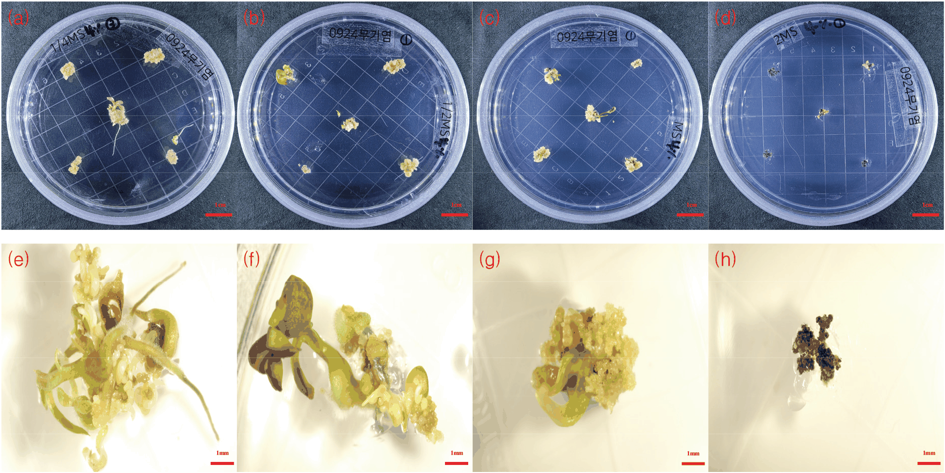
체세포배 유도는 무기염과 탄소원 농도에 따라 유의한 차이를 나타냈다. 특히 무기염 농도가 체세포배 형성에 큰 영향을 미쳤으며, 배양 2주 차부터 배발생 캘러스가 증식하기 시작하였다. 낮은 무기염 농도에서는 구형(globular stage) 체세포배와 식물체 형성이 동시에 진행되는 양상이 관찰되었다(Figure 4). 2배 농도의 MS 배지에서는 대부분의 캘러스가 갈변하며 고사되었다[Figure 4(h)].
Figure 4.
Induction of somatic embryos according to inorganic salts strength of MS medium from embryogenic callus. (a, e) 1/4MS; (b, f) 1/2MS; (c, g) MS; (d, h) 2MS. Scale bars represent 1 cm(a-d) and 1 mm(e-h).
Download Original Figure
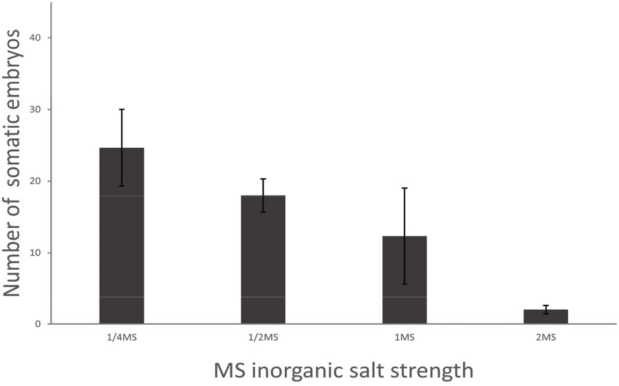
배양 6주 차 기준 체세포배 수를 분석한 결과, 1/4 MS 배지에서 24.7±5.4개로 가장 많은 체세포배가 형성되었고, 1/2 MS(18.0±2.3개), MS(12.3±6.7개), 2 MS(2.0±0.6개) 순으로 감소하였다(Table 5, Figure 5). 이는 낮은 무기염 농도가 배발생 캘러스 증식과 체세포배 형성에 유리함을 시사하며, 올소시폰(Orthosiphon aristatus) 연구(Sianipar and Mariska, 2020)와 유사한 경향을 보였다.
Table 5.
Effects of inorganic salts strength of MS medium on somatic embryo induction from leaf explants of D. spathulata.
| MS inorganic salts strength |
Embryogenic cell and somatic embryo induction (%) |
Mean number of somatic embryos/explant |
| 1/4MS |
86.7 |
24.7±5.4 |
| 1/2MS |
73.3 |
18.0±2.3 |
| MS |
46.7 |
12.3±6.7 |
| 2MS |
6.7 |
2.0±0.6 |
Download Excel Table
Figure 5.
Effects of inorganic salts strength on induction of somatic embryo from embryogenic callus. Each treatment consisted of 5 somatic embryos in a petri dish and was repeated three times. Error bars represent SE (N = 15).
Download Original Figure
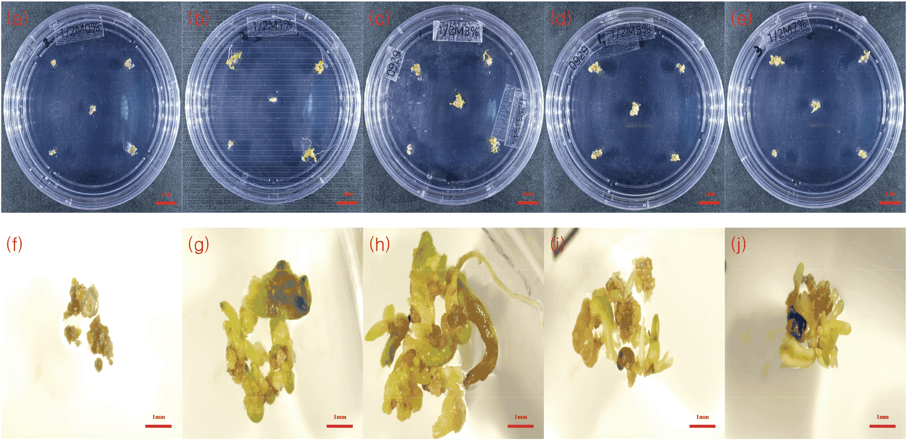
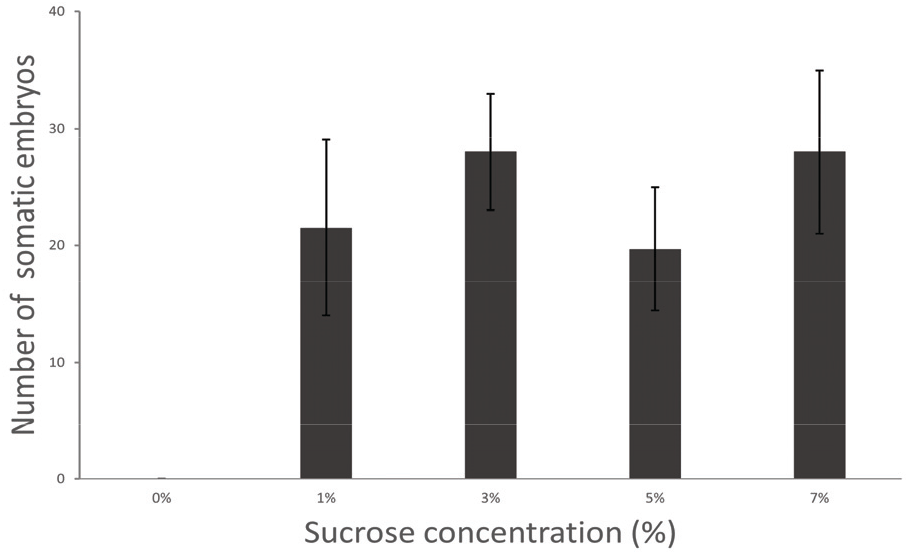
또한 탄소원으로 첨가된 Sucrose의 존재 여부가 체세포배 형성에 필수적임이 확인되었다. 체세포배 발달은 배양 4주 차부터 본격적으로 진행되었으며, Sucrose가 첨가되지 않은 배지에서는 체세포배 형성이 전혀 관찰되지 않았다. 반면, 3% sucrose 첨가 처리에서는 체세포배와 식물체 형성이 동시에 진행되는 것이 육안으로 확인되었다(Figure 6). 농도를 달리한 처리(1%, 3%, 5%, 7%)에서 각각 21.5±7.5개, 28.0±5.0개, 19.7±5.2개, 28.0±7.0개의 체세포배가 형성되었으나(Table 6), 수치상으로는 3%와 7% sucrose 첨가 시 체세포배 형성 수가 가장 많았으나, 일원 배치 분산분석 결과 농도 간의 통계적 유의차는 인정되지 않았다(Figure 7). 이는 sucrose의 존재 여부가 정량적 농도 차이보다 체세포배 유도에 더 큰 영향을 미침을 의미한다.
Figure 6.
Induction of somatic embryos according to the sucrose concentration from embryogenic callus. (a, f) 0%, (b, g) 1%, (c, h) 3%, (d, i) 5%, (e, j) 7% sucrose. Scale bars represent 1 cm(a-e) and 1 mm(f-j).
Download Original Figure
Table 6.
Effects of the sucrose concentration on somatic embryo induction from leaf explants of D. spathulata.
| Sucrose concentration |
Embryogenic cell and somatic embryo induction (%) |
Mean number of somatic embryos/explant |
| 0% |
- |
- |
| 1% |
53.3 |
21.5±7.5 |
| 3% |
66.7 |
28±5.0 |
| 5% |
100 |
19.7±5.2 |
| 7% |
46.7 |
28±7.0 |
Download Excel Table
Figure 7.
Effects of sucrose concentrations on induction of somatic embryo from embryogenic callus. Each treatment consisted of 5 somatic embryos in a petri dish and was repeated three times. Error bars represent SE (N = 15).
Download Original Figure
4. 활성탄 농도에 따른 체세포배 유래 식물체 재분화 반응
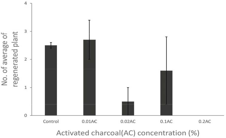
3번 실험에서 최적 조건으로 유도된 체세포배를 다양한 농도의 활성탄이 첨가된 배지에서 배양한 결과, 신초 출현 수에 차이가 관찰되었다. 이때 신초 출현은 체세포배로부터 새로운 줄기와 잎이 형성되어 식물체로 발달하기 시작하는 단계를 의미한다. 이는 활성탄이 배지 내 유리 페놀물질을 흡착하여 세포분화를 일부 유도한 결과로 해석될 수 있으나, 0.02% 이상의 농도에서는 신초 형성이 급격히 억제되었다. 이는 활성탄의 과도한 첨가가 페놀물질뿐만 아니라 배지 내 필수 영양소나 생장조절제까지 흡착하여 세포 발달에 부정적인 영향을 미친 것으로 판단된다. 활성탄을 첨가하지 않은 배지에서는 체세포배 당 평균 2.5±0.1개의 신초가 형성되었으며, 0.01% 활성탄 첨가 시 2.7±0.7개로 대조구 대비 미미하게 증가하였다. 그러나 0.02% 처리에서는 0.5±0.5개로 급격히 감소하였고, 0.2% 처리군에서는 신초가 전혀 형성되지 않고 소멸하였다(Figure 8). 이는 0.02% 이상의 활성탄 처리군이 신초 발달을 강력하게 저해함을 시사한다.
Figure 8.
Effects of Activated charcoal concentration(%) on the number of average of regenerated plant. Each treatment consisted of 5 somatic embryos in a petri dish and were repeated three times. Error bars represent SE (N = 15).
Download Original Figure
활성탄을 첨가하지 않거나 소량(0.01%) 첨가한 조건에서 신초 형성이 가장 양호하였으며, 0.02% 이상의 처리군에서는 발달이 저해되었다(Figure 9). 그러나 전반적으로 활성탄의 첨가 여부는 체세포배 유도 개수에는 유의한 영향을 미치지 않았으며, 신초 출현 수에도 제한적인 효과만을 보였다.
Figure 9.
Plant regeneration according to Activated charcoal concentration from somatic embryos. (a) 0%, (b) 0.01%, (c) 0.02%, (d) 0.1%, (e) 0.2% activated charcoal. Scale bars represent 1 cm.
Download Original Figure
5. 주걱댕강나무의 체세포배발생 및 식물체 재분화
주걱댕강나무의 잎 절편체 캘러스를 이용하여 체세포배발생을 통한 식물 재분화 체계를 확립하였다. 분화가 일어나는 유백색의 배발생 캘러스는 1/2MS 배지에서 원활하게 증식되었으며, 4주 간격으로 새로운 배지로 계대배양을 실시하였다.
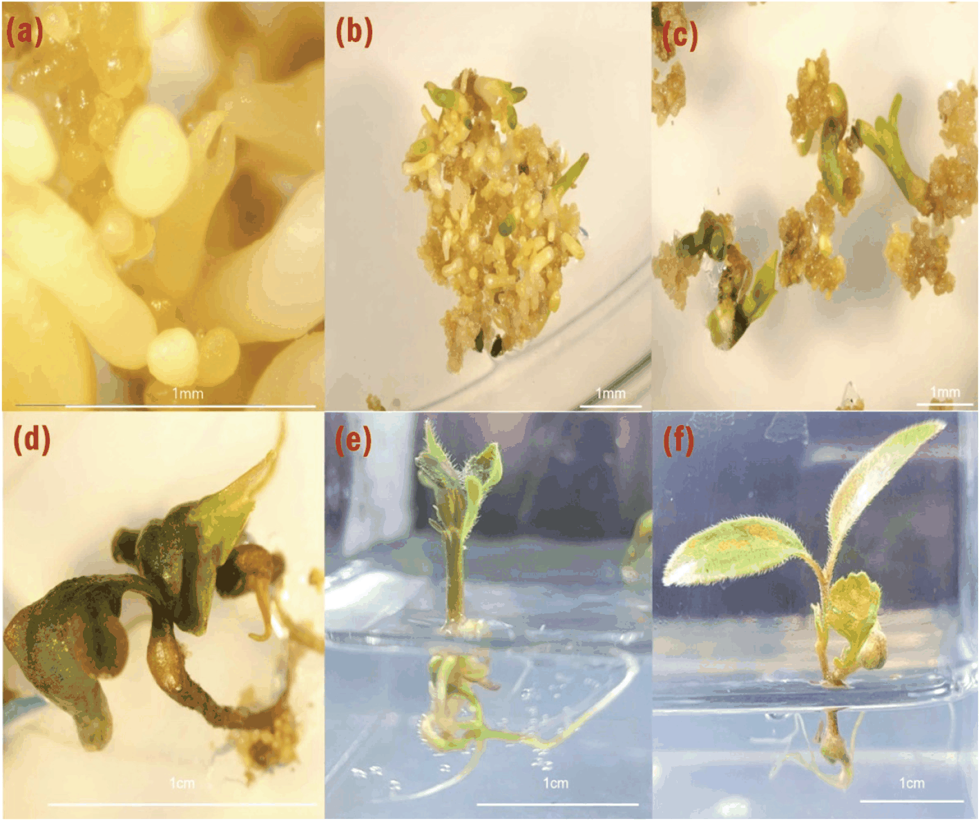
증식된 배발생 캘러스를 1.0 mg/L의 BAP가 첨가된 1/2MS 배지에서 배양한 결과, 체세포배는 기간이 긴 globular 단계에서[Figure 10(a)] heart 단계와 torpedo 단계를 동시에 거치며 자엽기 단계로 발달하였다[Figure 10(b)](Zimmerman, 1993).
Figure 10.
Somatic embryogenesis of D. spathulata. Leaf explants cultured on 1/2MS medium induced (a) globular and heart-shaped. Indirect embryogenic callus and globular - torpedo (b) stage embryo induced from leaf explant on 1/2MS medium supplemented BAP 1.0 ㎎/L after culture for 8 weeks. (c,d) Germinated somatic embryos were developed from leaf explants on MS medium supplemented BAP 1.0 ㎎/L after culture for 4 weeks. (e,f) Shoot and root formation on MS hormone free medium for 6 week. Scale bars represent 1 mm.
Download Original Figure
광조건에서 6주간 배양한 체세포배는 성공적으로 발아하였고[Figure 10(c, d)], 소식물체로 발달하였다[Figure 10(e, f)]. 이 결과는 주걱댕강나무에서도 체세포배 발생을 통한 전 식물체 재분화 체계가 가능함을 실험적으로 입증하였다.
결 론
주걱댕강나무의 잎절편을 대상으로 캘러스를 유도한 결과 BAP 1.0 mg/L와 NAA 2.0 mg/L 혼용 처리에서 캘러스 직경과 생중량이 통계적으로 가장 높은 값을 나타냈다. 일부 처리구에서는 비특이적 기관분화가 확인되었다. 또한 zeatin과 NAA 혼용 처리에서는 부정근 형성이 뚜렷하게 나타났으나 이는 체세포배 유도와는 무관한 것으로 판단되었다. BAP와 NAA의 5.0 mg/L 이상 고농도 처리구는 캘러스의 갈변 및 생장 저해를 초래하여 분석 대상에서 제외하였으며, 본 연구에서는 2.0 mg/L 이하의 적정 농도 결과를 중심으로 체세포배 중심의 재분화 체계를 확립하였다.
배발생 캘러스는 생장조절제를 첨가하지 않은 배지와 낮은 무기염 농도 배지에서 상대적으로 높은 빈도로 유도되었다. 체세포배 유도 실험에서는 1/4 MS 배지에서 체세포배 형성이 가장 많았으며, sucrose 3% 조건에서 체세포배 발달과 식물체 형성이 동시에 관찰되었다. 그러나 sucrose 농도 차이는 체세포배 유도 개수에 유의한 영향을 미치지 않았다. 활성탄 농도에 따른 체세포배 발아 실험에서는 0.01%의 저농도 활성탄 처리 또는 활성탄을 첨가하지 않은 처리구에서 식물체 분화가 상대적으로 높은 빈도로 나타났다.
이 연구는 체세포배발생 기술을 활용한 주걱댕강나무의 최초 엽배양 연구로, 희귀종 보전을 위한 실질적 가능성을 확인하였으며, 후속 연구를 통해 확립된 시스템을 바탕으로 대량증식 효율을 극대화할 필요가 있다. 특히, 다양한 환경 조건을 고려한 실험을 통해 현장 적용성을 검증하고, 기술의 응용 범위를 확대함으로써 희귀종 복원과 생물다양성 보전에 기여할 수 있을 것이다. 이 연구에서 제시된 엽배양 기술은 주걱댕강나무뿐 아니라 유사한 생태적 특성을 가진 희귀종에도 적용 가능성을 지닌다. 나아가 복원 생태학적 관점에서 이 기술은 멸종 위기 식물의 생존 가능성을 높이고, 생태계 복원과 건강성 유지에 중요한 과학적 도구로 활용될 것으로 기대된다.
감사의 글
본 연구는 정부(환경부)의 재원으로 국립생물자원관 ‘자생생물 대량증식 연구기반 구축’ 사업(과제번호: 2023년 NIBR202315101, 2024년 NIBR202414201, 2025년 NIBR202513201)과 산림청(한국임업진흥원) 산림과학기술 연구개발사업(RS-2024-00404388)의 지원으로 수행되었습니다.
연구 진행 과정에서 소중한 조언과 피드백을 제공해 주신 김창길 교수님께 깊은 감사를 드립니다.
References
Ahn, J.W. 2003. A study on the characteristics and propagation methods of Abelia mosanensis and Abelia grandiflora. MS Thesis, Department of Landscape Architecture, Sungkyunkwan Univ., Korea. pp. 1-49 (in Korean).

Ahn, C.H. and Lee, H.S. 2023. Regeneration of
Xanthoceras sorbifolium via Adventitious Shoot Formation from Immature Seeds. Agricultural Life Science Research 57(1): 45-50.

Ammirato, P.V. 1983. Embryogenesis. In: Evans, D.A., Sharp, W.R., Ammirato, P.V. and Yamada, Y. (eds.), Handbook of Plant Cell Culture, Vol. 1: Techniques for Propagation and Breeding. Macmillan Publishing Co., New York, 82-123.
Chu, Y.R., Park, S.H. and Jung, E.J. 2020. Propagation of endangered species, Daphne pseudomezereum var. koreana via in vitro bud culture. Journal of Korean Society of Forest Science 109(2): 189–194.
Dalila, Z., Dhiya, H.J., Jaafar, H. and Manaf, A.A. 2013. Effects of 2,4-D and kinetin on callus induction of Barringtonia racemosa leaf and endosperm explants in different types of basal media. Asian Journal of Plant Sciences 12(1): 21-27.
Dirr, M.A. 1977. Manual of Woody Landscape Plants: Their Identification, Ornamental Characteristics, Culture, Propagation and Uses. Stipes Publishing Co., Champaign, Illinois.
Duncan, D.B. 1955. Multiple range and multiple F tests. Biometrics 11(1): 1-42.
Im, H.J., Jeong, D.S., Lee, W.J., Kim, K.M., Song, S.H., Kim, D.W., Yang, J.C., Jung, J.C. and Yi, J.S. 2024. Influence of plant growth regulator and light-emitting diodes on formation and induction for callus of Styphnolobium japonicum (L.) Schott. Journal of Agriculture & Life Science 58(1): 143-151.
Jang, J.G. 2021. A study on conservation biology of Diabelia spathulata in Korea. PhD Dissertation, Kyungpook National University, Daegu, Korea.
Jeong, Y.M., Lee, H.J. and Kim, S.H. 2010. Numerical study on the impact of regional warming on the meteorological field and ozone concentration over the south-eastern part of the Korean Peninsula. Journal of Environmental Science International 19(12): 1431-1445.
Kim, T.J. 1996. Korean Resource Plants IV. Samsung Cultural Printing Co., Seoul, Korea.
Kim, T.Y. and Kim, J.S. 2018. Korean Trees. Dolbegae, Seoul, Korea.
Malik, S.I., Khan, M.A., Ahmad, S. and Ali, R. 2003. Effect of 2,4-dichlorophenoxyacetic acid on callus induction from mature wheat (Triticum aestivum L.) seeds. Int. J. Agric. Biol 6(1): 156-159.
Moon, H.K., Suk, G.Y., Kwon, Y.J. and Son, S.H. 1999. Micropropagation of a rare species, Abeliophyllum distichum Nakai, via axillary bud culture. Korean Journal of Plant Tissue Culture 26(2): 133.
Moon, H.K. and Kim, Y.W. 2008. In vitro propagation of a rare and endangered species, Echinosophora koreensis Nakai, by axillary bud culture. Journal of Plant Biotechnology 35(3): 229-234.
Murashige, T. and Skoog, F. 1962. A revised medium for rapid growth and bioassays with tobacco tissue cultures. Physiologia Plantarum 15(3): 473-497.
KNA (Korea National Arboretum). 2021. The National Red List of Vascular Plants in Korea. Korea National Arboretum, Pocheon, Korea.
Park, J.Y., Lee, S.H., Kim, D.S., Kong, H.J., Nam, G.H., Park, J.E. and Kang, J.W. 2024. Optimal concentrations of plant growth regulators and cultivation conditions for plant regeneration from leaf explants of Aster maackii. Journal of Plant Biotechnology 51(1): 286-293.
Phua, Q.Y., Chin, C.K., Asri, Z.R.M., Lam, D.Y.A., Subramaniam, S. and Chew, B.L. 2016. The callugenic effects of 2,4-dichlorophenoxy acetic acid (2,4-D) on leaf explants of Sabah snake grass (Clinacanthus nutans). Pakistan Journal of Botany 48(2): 561-566.
Pinto, G., Valentim, H., Costa, A., Castro, S. and Santos, C. 2002. Somatic embryogenesis in leaf callus from a mature Quercus suber L. tree. In Vitro Cellular & Developmental Biology – Plant 38(6): 569-572.
Sianipar, N.F. and Mariska, I. 2020. Micropropagation of Orthosiphon aristatus through indirect and direct organogenesis. Jurnal Teknologi (Sciences & Engineering) 82(3).
Tahir, S.M., Victor, K. and Abdulkadir, S. 2011. The effect of 2,4‑dichlorophenoxy acetic acid (2,4‑D) concentration on callus induction in sugarcane (Saccharum officinarum). Nigerian Journal of Basic and Applied Sciences 19(2): 213-217.
Williams, E.G. and Maheswaran, G. 1986. Somatic embryogenesis: factors influencing coordinated behaviour of cells as an embryogenic group. Annals of Botany 57(4): 443-462.
Yi, M.H., Yoo, S.T. and Jang, J.G. 2021. Vegetation structure and growth environment of Diabelia spathulata (Siebold & Zucc.) Landrein population in Mt. Cheonseong, Korea. Korean Journal of Plant Resources 34(4): 346-361.

 PDF Download
 PubReader
 Export Citation
 Email the Author
 Share Facebook
 Share Twitter
 Metrics (View:460)